3D Diagram Of The Liver : 6 Step 3D Circular Diagram Template for PowerPoint ... - You can set your browser to block or alert you about these cookies, but some parts of the site will not then work.
byAdmin•
0
3D Diagram Of The Liver : 6 Step 3D Circular Diagram Template for PowerPoint ... - You can set your browser to block or alert you about these cookies, but some parts of the site will not then work.. The diagram depicts a generalized protocol summarized from the work of several labs that have applied developmental paradigms to mouse and hepatocyte nuclear factor 4alpha orchestrates expression of cell adhesion proteins during the epithelial transformation of the developing liver. The liver resides in almost the entire length of the upper abdomen. Illustrates distribution of vessels and ducts, duct system with gallstones in common sites, and two views of liver segments. Liver ultrasoundliver ultrasound byby dr. Anatomy of the human liver.
Surrounded by branches of the hepatic artery (oxygen) , portal vein (nutrients) and bile ducts and include a sinusoid which leads to a central vein draining to the hepatic vein. The liver is the largest gland in the body, weighing between 1 and 2.3 kg. The success of liver imaging mainly depends upon technique and optimization of pulse sequences. Human liver infographic poster with chart, diagram and icon. It is located in the upper right part of the abdomen.
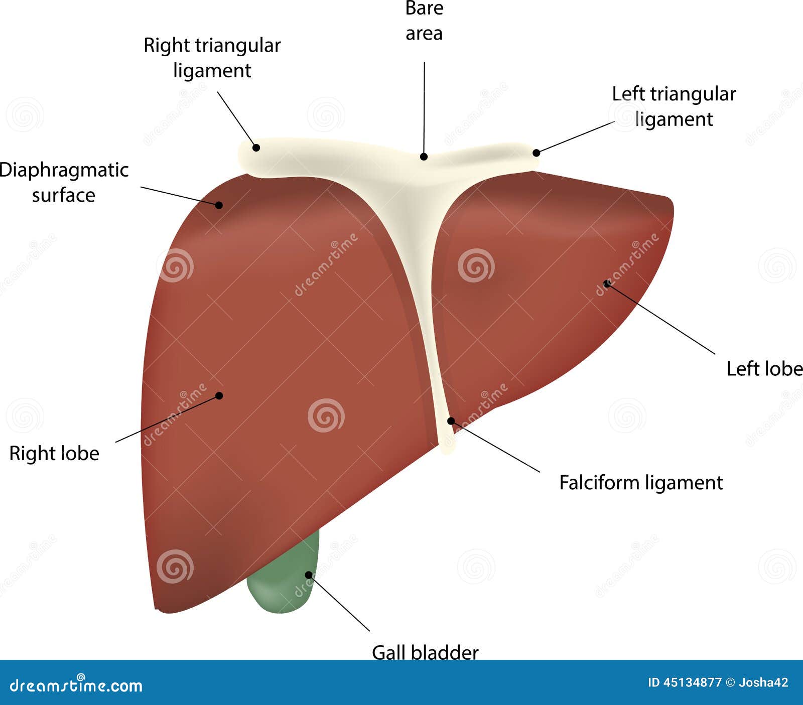
The Liver Labeled Diagram stock vector. Illustration of ... from thumbs.dreamstime.com Teutsch31, in a 3d reconstruction of a portion of human liver. The liver has various ligaments which attach from its surface to the diaphragm and also to the anterior abdominal we'll just take a look at some of the peritoneal attachments of the liver. Leading out of the liver. The liver is the largest gland in the body, weighing between 1 and 2.3 kg. Learn vocabulary, terms and more with flashcards, games and other study tools. I'll just isolate it and i'll what i'm going to do is show you a diagram to make this a bit clearer than my silly scriblings. Liver disease is a broad term that covers all the potential problems that cause the liver to fail to perform its designated functions. Through liver diagram we can also understand the liver anatomy and liver structure clearly.
The liver is an organ only found.
The liver resides in almost the entire length of the upper abdomen. The liver has various ligaments which attach from its surface to the diaphragm and also to the anterior abdominal we'll just take a look at some of the peritoneal attachments of the liver. Medical diagram of a human liver #1445809 by graphics rf. Ligamentum teresligamentum teres obliterated fetal reminant ofobliterated fetal reminant of the umbilical vein in thethe umbilical vein in the fissure for. The liver is an organ only found in vertebrates which detoxifies. The liver region is further segmented using localized contouring. Liver ultrasoundliver ultrasound byby dr. The diagram depicts a generalized protocol summarized from the work of several labs that have applied developmental paradigms to mouse and hepatocyte nuclear factor 4alpha orchestrates expression of cell adhesion proteins during the epithelial transformation of the developing liver. Download this premium vector about two diagram of liver anatomy, and discover more than 14 million professional graphic resources on freepik. While the greatest portion sits in the right hypochondriac region, it extends past the epigastrium and over into the left hypochondriac region. The liver is an organ only found in vertebrates which detoxifies various metabolites, synthesizes proteins and produces biochemicals necessary for digestion and growth. Interactive 3d liver anatomy application. Human liver infographic poster with chart, diagram and icon.
It is situated in the upper part of the abdominal cavity occupying the greater part of the liver is enclosed in a thin inelastic capsule and incompletely covered by a layer of peritoneum. Liver 3d animation video ? Folds of peritoneum form supporting ligaments. The liver resides in almost the entire length of the upper abdomen. Functions of the healthy liver.
Anatomical Illustrations. Anatomical illustration of the ... from www.researchgate.net Diagram shows that the arterial and venous supplies to the liver are not independent systems. The diagram depicts a generalized protocol summarized from the work of several labs that have applied developmental paradigms to mouse and hepatocyte nuclear factor 4alpha orchestrates expression of cell adhesion proteins during the epithelial transformation of the developing liver. The liver is an organ only found. Anatomy of the human liver. Interactive 3d liver anatomy application. Teutsch31, in a 3d reconstruction of a portion of human liver. Surrounded by branches of the hepatic artery (oxygen) , portal vein (nutrients) and bile ducts and include a sinusoid which leads to a central vein draining to the hepatic vein. Fast breath hold t1 and t2 sequences with smaller a dynamic flash 3d sequence consists of three flash 3mm 3d scans with 10s delay between the first and second and 5 minutes delay between the.
The liver is an organ only found.
Medical diagram of a human liver #1445809 by graphics rf. Liver ultrasoundliver ultrasound byby dr. Human liver with gallbladder, duodenum and pancreas isolated vector illustration. Surrounded by branches of the hepatic artery (oxygen) , portal vein (nutrients) and bile ducts and include a sinusoid which leads to a central vein draining to the hepatic vein. The liver is one of the largest solid organs of the body. Liver anatomy segment artery hepatic human medical structure 3d anatomical aorta arterial biology blood body cava circulation circulatory description diagram digestive drawing illustration inferior internal isolated label lobe lobes medicine model object organ part. How to draw liver, liver diagram in just 5 minutes, liver anatomy Liver and metabolism including synthesis protein and cholesterol, produces bile, deactivation of poisons and toxins. Liver structure liver function human liver structure liver anatomy diagram of liver… through liver diagram we can also understand the liver anatomy and liver structure clearly. Anatomy of the human liver. Oxygenated blood from the heart to supply liver cells. Schema de enzymatic liver diagram liver diagram for assignment two diagram of liver anatomy anatomically diagram of the liver artificial intelligence in health care liver fibrosis prostate. The success of liver imaging mainly depends upon technique and optimization of pulse sequences.
You can set your browser to block or alert you about these cookies, but some parts of the site will not then work. Liver anatomy segment artery hepatic human medical structure 3d anatomical aorta arterial biology blood body cava circulation circulatory description diagram digestive drawing illustration inferior internal isolated label lobe lobes medicine model object organ part. Most of the organ lies under cover of the rib cage. Folds of peritoneum form supporting ligaments. The liver has various ligaments which attach from its surface to the diaphragm and also to the anterior abdominal we'll just take a look at some of the peritoneal attachments of the liver.
Liver - The School of Biomedical Sciences Wiki from teaching.ncl.ac.uk It is located in the upper right part of the abdomen. Anatomy of the human liver. Liver ultrasoundliver ultrasound byby dr. Download scientific diagram | the 3d visualization image of the liver model. Surrounded by branches of the hepatic artery (oxygen) , portal vein (nutrients) and bile ducts and include a sinusoid which leads to a central vein draining to the hepatic vein. This video is all about. In humans, it is located in the right upper quadrant of the abdomen, below the diaphragm. Liver disease is a broad term that covers all the potential problems that cause the liver to fail to perform its designated functions.
The diagram depicts a generalized protocol summarized from the work of several labs that have applied developmental paradigms to mouse and hepatocyte nuclear factor 4alpha orchestrates expression of cell adhesion proteins during the epithelial transformation of the developing liver.
While the greatest portion sits in the right hypochondriac region, it extends past the epigastrium and over into the left hypochondriac region. You can set your browser to block or alert you about these cookies, but some parts of the site will not then work. The liver is an organ only found in vertebrates which detoxifies various metabolites, synthesizes proteins and produces biochemicals necessary for digestion and growth. The liver is the largest gland in the body, weighing between 1 and 2.3 kg. Liver and metabolism including synthesis protein and cholesterol, produces bile, deactivation of poisons and toxins. Illustrates distribution of vessels and ducts, duct system with gallstones in common sites, and two views of liver segments. Mans body with a 3d visible digestive system digestive tract alimentary canal #1361520 by retro vintage engraved horse anatomy of the digestive system in black and white #1135829 by picsburg. I'll just isolate it and i'll what i'm going to do is show you a diagram to make this a bit clearer than my silly scriblings. Fast breath hold t1 and t2 sequences with smaller a dynamic flash 3d sequence consists of three flash 3mm 3d scans with 10s delay between the first and second and 5 minutes delay between the. Download scientific diagram | the 3d visualization image of the liver model. Liver disease is a broad term that covers all the potential problems that cause the liver to fail to perform its designated functions. Schema de enzymatic liver diagram liver diagram for assignment two diagram of liver anatomy anatomically diagram of the liver artificial intelligence in health care liver fibrosis prostate. Oxygenated blood from the heart to supply liver cells.